The aim of the Multiple Molecular Imaging Research Laboratory is engaged in research on metals in biology. In order to achieve the better understandings of roles, functions and regulation of metals in biological systems, we developed the multitracer technology and the multiple molecular imaging systems which is using by semiconductor Compton telescope. The Multiple Molecular Imaging Research Laboratory performs following researches: (1) Developments of the multitracer technology by using accelerator facilities. (2) Application studies of the multitracer technology in the fields of chemistry, biology, medicine, pharmacy, nutrition, environment, agriculture, etc. (3) Developments of the Gamma Ray Emission Imaging as a tool for the multi-molecular imaging apparatus (4) Basic research for drug discovery for use the multi-molecular imaging. (5) Researches of biological and physiological function of bio-trace elements by using the new technology

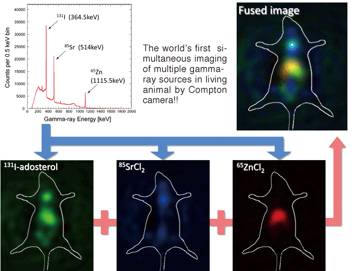
Multiple Molecular Imaging Research Laboratory is engaged in research on metals in biology. In order to understand the roles, functions, and regulation of metals in biological systems, we are developing a new gamma ray imaging system called GREI (Gamma-Ray Emission Imaging) by applying principle of Compton camera to divided-electrode germanium semiconductor. Using this system, we have succeeded in obtaining the world's first simultaneous multiple molecular image in living animal, and showed it is applicable to life sciences research such as metabolic physiology and molecular biological study in trace element.
We strongly believe simultaneous multiple molecular imaging that GREI will produce is an effective tool for clinically crucial early diagnosis of intractable disease such as cancer and metabolic disease, and evaluation of transplant and regenerative treatment. Currently, we are working to achieve high sensitivity and resolution of the system, and discovering new multiple molecular imaging probes to establish a clinically applicable new generation image diagnosis system.
【Development of Multiple Molecular Imaging Methodology】
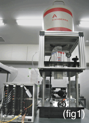
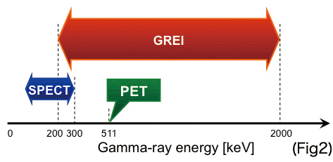
Factors involved in cancer and lifestyle related diseases are known to be complex. Information on several biological molecules is vital for advanced and precise diagnosis. However, positron emission tomography (PET) is only capable of tracing single molecule at a time, and single photon emission computed tomography (SPECT) can handle limited number of nuclei because it is only applicable for low-energy gamma rays. RIKEN is currently developing a semiconductor Compton camera called GREI (Gamma-Ray Emission Imaging) for practical usage (Fig 1). It can handle far wider energy range of gamma ray than any existing image diagnosis system and analyze multiple gamma rays simultaneously (Fig 2). By analyzing dynamics of multiple molecules simultaneously, it can alter the utility of molecular imaging and offer unprecedented diagnosis.
GREI is equipped with high-purity germanium semiconductor sensor, which detects position and energy of incident gamma rays with high sensitivity, and uses Compton scattering kinematics to determine the spatial distribution of gamma-ray sources. Furthermore, this system has high energy resolution, which can discriminate each molecule clearly by its gamma ray energy. Fig 3 shows simultaneous image of 65Zn accumulating around liver, 85Sr around spine and 131I around adrenal gland and thyroid gland. As shown, GREI can visualize simultaneous multiple molecular image by labeling each molecule with different energy source.
![Search[Google]](../common/image/hmenu_search.png)

![CMIS[RIKEN Center for Molecular Imaging Science]RIKEN Kobe Institute Center for Molecular Imaging Science](../common/image/hlogo_txt.png)


![[Multiple Molecular Imaging Research Laboratory] Team Leader: Shuichi Enomoto](../image/laboratory/lob06_titleimage.jpg)