![Search[Google]](../../common/image/hmenu_search.png)

![CMIS[RIKEN Center for Molecular Imaging Science]RIKEN Kobe Institute Center for Molecular Imaging Science](../../common/image/hlogo_txt.png)


The cerebrovascular disorder consists of a cerebral hemorrhage caused by a ruptured blood vessel, and a cerebral infarction caused by a clogged vessel. These diseases are the top two causes for people in Japan who require nursing care. Under normal conditions, the brain maintains a constant blood flow by its autoregulatory function which constricts or dilates a blood vessel, depending on alteration in blood pressure. However, researches reveal that the cerebrovascular disorder causes excessive production of biomolecules, which will rather exacerbate the symptoms of the disease than allow them to fulfill their autoregulatory function.
Experiments show that 20-Hydroxyeicosatetraenoc acid (20-HETE), presumably playing an autoregulatory function for cerebral blood flow, is transiently and abundantly produced in the rats when they have stroke. Whereas 20-HETE is thought to be one of the molecules which promote vascularization, and thought to help damaged blood vessels regenerate, it may be exacerbating initial symptoms of a cerebral infarction, because of its another function of blood vessel constriction. In fact, it is confirmed that inhibition of the synthesis of 20-HETE for animal cerebral infarction models effectively prevents expansion of affected regions. The cause of the production of 20-HETE is believed to be a transiently elevated activity of 20-HETE synthetic enzyme in the brain, but the details of the mechanism are still unclear. A clear understanding of the relation between brain disorders and 20-HETE synthetic enzyme would provide critical information to understand the disease states and prognosis. This time, our research team has developed a PET probe [11C] TROA targeting 20-HETE synthetic enzyme and successfully performed the live-imaging of it.
[11C] TROA is a 20-HETE inhibitor of activity N' (4-Dimethylaminohexyloxy) phenyl imidazole (TROA) labeled by a 11C radioisotope using high performance C-[11C] methyl reaction. The PET exam using normal rats showed high concentration of [11C] TROA on kidneys, livers and other organs which are known to have abundant 20-HETE synthetic enzyme under normal conditions. On rat cerebral infarction models,[11C] TROA was recognized to highly concentrate in the affected sphere of the brain, peaking on day 7 after strokes. This indicates a transient elevation of the activity of 20-HETE synthetic enzyme following cerebral infarctions. The fact was confirmed by measuring the contents of RT-PCR and 20-HETE as well as by PET exams.
Our study results showed that changes in the activity of a 20-HETE synthetic enzyme are observable by the PET exam and such observation will serve as a possible critical diagnostic method to establish the most suitable treatment strategy. We are going to perform PET imaging of [11C] TROA on primate models to explore applicability of this technique to humans in the future. Furthermore we will pursue possibility of the 20-HETE synthetic enzyme inhibitor as therapeutic drugs to relieve the symptoms.
* This study was a joint program between a study group in Graduate School of Pharmaceutical Sciences, Osaka University, and Taisho Pharmaceutical Co., Ltd, and teams in Functional Probe Research Laboratory (team leader Hirotaka Onoe, researcher Toshiyuki Kawasaki) and Molecular Imaging Medicinal Chemistry Laboratory (team leader Masaaki Suzuki, researcher Keiko Shirakami) and Molecular Imaging Labeling Chemistry Laboratory (team leader Hisashi Doi, technical staff Tomoko Mori), RIKEN Center for Molecular Imaging Science.
See here for more details of the study.
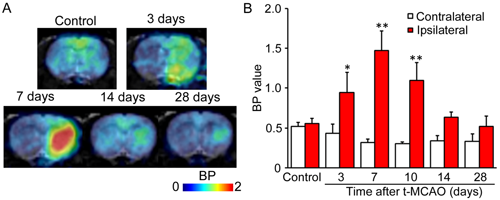 |
| Changes in the activity of a 20-HETE synthetic enzyme on rat cerebral infarction models. A) In the brain of a rat with occluded middle cerebral artery (on the right side), we observed the volume of [11C] TROA binding on 20-HETE synthetic enzyme using PET imaging. The red signal indicates that the high degree of binding reached a peak on day 7 after operation. B) The graph of the results of experiments using three individuals. Based on statics, the signal for [11C] TROA in the ipsilateral region is significantly higher than the signal for that in the corresponding contralateral region. |
