![Search[Google]](../../common/image/hmenu_search.png)

![CMIS[RIKEN Center for Molecular Imaging Science]RIKEN Kobe Institute Center for Molecular Imaging Science](../../common/image/hlogo_txt.png)


Early detection of glaucoma by PET imaging
-New diagnostic technique to examine ocular diseases in brain-
Glaucoma is the leading cause of blindness in Japan. It is among highly frequent diseases of eye and 5% of adults at the age of 40 or older are known to suffer from this disease. The initial symptom of glaucoma is the appearance of invisible areas called "scotoma" within the visual field. However, it usually takes a long time before people are aware of the presence of scotoma in their own visual field because the scotoma spread gradually from the very periphery of the visual field. Moreover, they are often devoid of subjective symptoms such as pain or itching of eyes. Such an asymptomatic period will often delay the diagnosis and result in progression of the disease and even fatal blindness. Only less than 20% of the glaucoma patients receive appropriate treatment at a clinic.
The cause of glaucoma is mostly due to the elevated pressure of inner eye compartment (intraocular pressure), which gradually damages optic nerves or retina deep in the eye. Thus, measurement of intraocular pressure may help to specifically diagnose this disease at the initial stage. In some cases, however, similar pathological conditions develop with scotoma even though the intraocular pressure is within a normal range. Thus, for more accurate diagnosis, the doctors have needed not only measurement of intraocular pressure but also other measurement devices for finding the glaucoma pathology sensitively. Moreover, it has been suggested that glaucoma affects not only eye and optic nerve but also a certain par of brain involved in processing information from the eyes. Therefore, a new way to definitely diagnose glaucoma in its early stage has long been awaited.
By using a molecular imaging technique, our research team investigated what actually happens in the brains of glaucoma using a glaucoma model monkey. Using positron emission tomography (PET), they focused on finding the inflammatory reaction, particularly, activated microglia, which is known as one of important immune cells in the brain. PET imaging in the glaucomatous monkey with its early-stage revealed that microglia-associated PET probes were accumulated in the deep in the brain, at the lateral geniculate body, where the optic nerves from the eyes communicate with cerebral neurons through the inter-neuronal gap, "synapses". This microglial activation was also accompanied by shrinkage and degeneration of neurons in the lateral geniculate body in a histopathological examination. These results suggest the possibility that glaucoma, which has been considered to be a disease of eye, could be correctly diagnosed by brain PET imaging at the initial stage of the disease. The research team is now planning to proceed with the clinical study in order to determine whether this screening method can be applied to glaucoma patients as a non-invasive diagnostic tool for early-stage glaucoma.
* This study was a joint program between a study group in Department of Biofunctional Evaluation, Gifu Pharmaceutical University (Hideaki Hara, Masamitsu Shimazawa, Yasushi Ito et al.), Functional Probe Research Laboratory, RIKEN Center for Molecular Imaging Science (director Yasuyoshi Watanabe) (team leader Hirotaka Onoe, sub-team leader Takuya Hayashi, research associate Hajime Yamanaka), and others.
* A part of this study was subsidized by the Ministry of Education, Culture, Sports, Science and Technology under "Molecular imaging research program," a commissioned program conducted from 2005 to 2009.
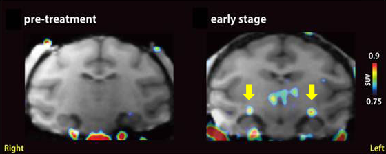 |
| PET images of glaucoma model monkey. Live imaging was conducted to visualize activated microglia using 11C-PK11195 as a PET probe. The lateral geniculate body of the brain is shown in the coronal plane of the head. PET probe signals of 11C-PK11195 were not detected in the brain before inducing glaucoma (left), but were found in the lateral geniculate bodies in the early stage of glaucoma (yellow arrow in right). |
